


The anus is the opening at the end of the digestive tract where stool leaves the body.
The rectum is the section of the digestive tract above the anus where stool is held before it passes out of the body through the anus.
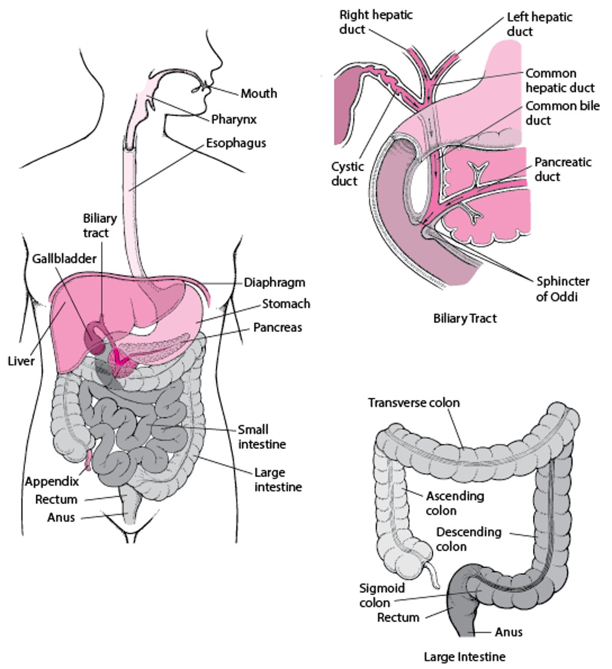
The Digestive System

The anus is formed partly from the surface layers of the body, including the skin, and partly from the intestine.
The rectal lining consists of glistening red tissue containing mucus glands—much like the rest of the intestinal lining. The lining of the rectum is relatively insensitive to pain, but the nerves from the anus and nearby external skin are very sensitive to pain.
The veins from the rectum and anus drain mostly into the portal vein, which leads to the liver, and then into the general circulation. Some of these veins drain directly into the pelvic veins and then into the general circulation. The lymph vessels of the rectum drain into lymph nodes in the lower abdomen. The lymph vessels of the anus drain into the lymph nodes in the groin.
A muscular ring (anal sphincter) keeps the anus closed. This sphincter is controlled subconsciously by the autonomic nervous system. However, part of the sphincter can be relaxed or tightened at will.
Disorders of the anus and rectum include
Diagnosis of Anal and Rectal Disorders
A doctor's evaluation
Anoscopy or sigmoidoscopy
Possibly computed tomography (CT) scan or magnetic resonance imaging (MRI)
To diagnose disorders of the anus and rectum, a doctor inspects the skin around the anus for any abnormality. With a gloved finger, the doctor probes the rectum. For women, this is often done along with a manual examination of the vagina (see Gynecologic Examination). Doctors often also examine the abdomen.
Next, a doctor looks into the anus and rectum with a 3- to 10-inch (about 7- to 25-centimeter) rigid viewing tube (anoscope or proctoscope). A longer, flexible tube (sigmoidoscope) may then be inserted so that the doctor can observe as much as 2 or more feet of the large intestine.
An anoscopy or sigmoidoscopy (see Endoscopyanesthetic before proceeding with the examination. Sometimes a cleansing enema to rid the lower part of the large intestine of stool is given before sigmoidoscopy.
Tissue and stool samples for microscopic examination and cultures may be obtained during sigmoidoscopy.


